


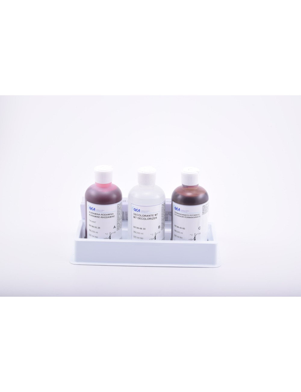
TB AURAMINE-RHODAMINE (TRUANT) KIT, 3 x 250 mL
999320
To access the documentation you must be registered Register
For invitro diagnostic.
| State | Liquid |
| Storage temp. | +10 / +35 ºC |
| Technique | Fluorescent staining |
Documentation
 Download
Download
Download
Download
Download
Previous batches :
Download
PRINCIPLE
Mycobacteria are acid-fast microorganisms that have a special lipid cell wall, which contains mycolic acid, amongst other substances. This enables the wall to resist destaining with acid-alcohol after staining with basic dyes such as fuchsin. The cell wall appears a reddish-pink colour, while the remainder of the microorganism is stained with the counterstain, normally Methylene Blue. The technique for detecting acid-fast microorganisms by fl uorescence is similar to the Ziehl classic staining method. The fenicade fuchsine is replaced by a fl uorescent dye with added phenol - in this case Auramine-Rhodamine, according to the Truant technique. The Auramine-Rhodamine binds to the mycolic acids of the wall and the free dye is removed by rinsing with the acidic destaining agent. The counterstain, Potassium Permanganate or Thiazine Red, removes the non-specifi c background fl uorescence. Compared to the classic staining methods, fl uorescence staining provides the advantage of greater visibility of the fl uorescent microorganisms on a dark background, making it easier to work with lower magnifi cation lenses, increasing the fi eld of vision and reducing the time required to evaluate the preparation.
DIAGNOSTIC USE
Stains for Auramine-rhodamine staining are used to perform the staining of cultures or samples which are suspected of containing mycobacteria, for early diagnosis of mycobacterial infection and the characterisation thereof.
For invitro diagnostic.
| State | Liquid |
| Storage temp. | +10 / +35 ºC |
| Technique | Fluorescent staining |