Menu

To access the documentation you must be registered Register


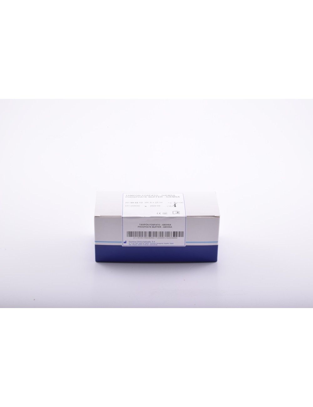
For Giemsa dilution. pH 7.2
| Storage temperature | +10 / +35 ºC |
| Estate | Liquid |
| Technique | Modified Romanowsky staining |
PRINCIPLE
The Giemsa stain belongs to the Romanowsky group of stains. It is named after Gustav Giemsa, the German chemist and bacteriologist who developed it. Romanowsky stains consist of Methylene Blue and its oxidised derivatives, basic dyes and the acidic dye, Eosin. Basic dyes bind to the acidic components of cells, nucleic acids, granules in neutrophils and acidic proteins which are stained a relatively deep red-purple colour, while eosin binds to haemoglobin, basic components of cell structures and eosinophil granules. The balance between Methylene Blue and its oxidised derivatives, and between these and Eosin, provides an essentially blue shade and a greater or lesser staining intensity, which are characteristics of each type of stain - Giemsa, May-Grünwald or Wright. This stain may be used alone or combined with the May-Grünwald stain. Its use allows the diff erential staining of blood cells. Some authors use the Giemsa stain or the May-Grünwald-Giemsa combination for the staining of parasites in blood.
INTENDED USE
Family of in vitro diagnostic medical devices intended for the morphological diff erentiation of nuclei and cytoplasm of platelets, white blood cells and red blood cells for hematologic and histologic assays. They are qualitative stains that provide information on the physiological or pathological state of the tissue sample when the staining obtained is interpreted by a trained professional. To obtain a diagnosis, the result is utilized alongside other information such as the patient’s medical history, physical status as well as other clinical and laboratory data obtained. For trained and specialized personne